
I encourage patients to come in early when they discover that they are pregnant, but I accept patients at almost anytime during the first two trimesters of pregnancy. An ultrasound is usually done on the first visit to confirm the due date. Typical appointments are every four weeks up to seven months, then every two weeks until your last month, when appointments are weekly. I perform all the standard and optional prenatal testing which is consistent among all doctors in Memphis. I do answer my own calls after hours as opposed to having a nurse on call. I deliver almost all of my own patients at either Baptist Hospital for Women or Methodist Germantown.
Your Prenatal Visits
At your first visit, we will review your medical history and usually perform an ultrasound to confirm your estimated due date.
How Often Will I Have Appointments?
- Until 28 weeks: Every four weeks
- 28–36 weeks: Every two weeks
- 36 weeks until delivery: Weekly appointments
Personalized Care
One of the advantages of my practice is continuity of care. When you call after hours, you speak directly with me rather than an answering nurse.
I also deliver nearly all of my own patients at Baptist Memorial Hospital for Women or Methodist Germantown Hospital.
Routine Prenatal Testing
First Prenatal Visit
Medical history, physical examination, blood type, Rh factor, anemia screening, infectious disease testing, and urine culture.
10 Weeks — NIPT
Noninvasive prenatal testing screens for Down syndrome, Trisomy 18, Trisomy 13, and certain sex chromosome abnormalities. It can also determine your baby's sex and Rh type if your Rh type is negative.
15–21 Weeks — AFP Screening
A maternal blood test that screens for open neural tube defects, including spina bifida.
18–22 Weeks — Anatomy Ultrasound
A detailed examination of your baby's developing organs, including the brain, spine, heart, kidneys, and extremities.
Additional Ultrasounds During Pregnancy
Seeing your baby on ultrasound is one of the most exciting parts of pregnancy. Although it is always enjoyable to watch your baby move and grow, ultrasounds in my office are performed for medical reasons rather than simply to obtain photographs.
Some pregnancies require additional ultrasounds during the third trimester. These may be recommended for women with chronic hypertension, diabetes, certain other medical conditions, or concerns about the baby's growth. Maternal age and other individual risk factors may also influence the need for additional monitoring.
If your baby appears smaller or larger than expected, an ultrasound may be performed to estimate fetal weight. Although ultrasound is helpful in evaluating growth, estimated fetal weights are not exact and can differ from the baby's actual birth weight by 10% or more.
For parents who would like additional keepsake images, several independent ultrasound studios offer elective 3D and 4D ultrasound photographs and video recordings. These are intended for entertainment rather than medical evaluation and should never replace a diagnostic ultrasound or routine prenatal care. The FDA discourages ultrasound examinations performed solely for keepsake images.
Additional Prenatal Testing
24–28 Weeks — Gestational Diabetes
A one-hour glucose screening test checks for gestational diabetes. You will drink a solution containing 50 grams of glucose and have your blood sugar checked one hour later.
28 Weeks — Rhogam
Patients with Rh-negative blood may need Rhogam to prevent the development of antibodies against Rh-positive fetal blood cells. If you had an NIPT drawn you will not need Rhogam if your baby is Rh negative.
36–37 Weeks — Group B Strep
A vaginal and rectal swab checks for Group B streptococcus. If positive, antibiotics during labor greatly reduce the risk of infection in your newborn.
Near Your Due Date
Cervical examinations may be offered to evaluate dilation and effacement as your due date approaches.
What If My Water Breaks?
Your water breaking may cause a sudden gush of fluid or a slow, continuous trickle. It can sometimes be difficult to distinguish amniotic fluid from urine or vaginal discharge.
If you believe your water has broken, contact me and go to the hospital for evaluation, even if you are not having contractions.
Eating Fish During Pregnancy
Fish provides protein and omega-3 fatty acids that support your baby's development. However, certain fish contain high levels of mercury.
Fish to Avoid
- King mackerel
- Shark
- Swordfish
- Tilefish from the Gulf of Mexico
- Orange roughy
- Bigeye tuna
- Marlin
Good Fish Choices
- Salmon
- Shrimp
- Catfish
- Crawfish
- Tilapia
- Trout
- Canned light tuna
The FDA recommends 8–12 ounces per week of a variety of lower-mercury seafood during pregnancy.

Why Nutrition is Important in Pregnancy
Good nutrition before, during, and after pregnancy supports the health of both mother and baby. Focus on the quality of your food rather than simply eating larger portions. Choose a variety of vegetables, fruits, beans, whole grains, nuts, seeds, and fish, along with healthy fats. Limit highly processed foods, fatty red meats, sweets, and sugary drinks. Your weight before pregnancy and your weight gain during pregnancy both matter. Being underweight, having obesity, or gaining too little or too much can increase the risk of complications. Addressing nutrition early—ideally before conception—can help support a healthier pregnancy. Avoid restrictive diets that eliminate major food groups, and discuss your nutritional needs and supplements with your healthcare provider. After delivery, a balanced diet continues to support recovery and breastfeeding. Breast milk provides nutrition tailored to your baby and is associated with health benefits for both mother and child.
Ten Things to Know
1. Healthy nutrition benefits mother and baby.
Improving nutrition and overall health before and during pregnancy supports fetal growth, healthier pregnancy outcomes, and potentially better lifelong health.
2. Nutrition influences a baby’s growth.
2. Nutrition influences a baby’s growth. Poor maternal nutrition can contribute to babies being too small or too large. These growth patterns are linked to a greater risk of chronic health problems later in life.
3. Pregnant teenagers need additional support.
Adolescents are still growing themselves, making good nutrition especially important. Professional guidance and practical support can help young mothers meet their nutritional needs before, during, and after pregnancy.
4. Healthy eating may reduce pregnancy complications.
A nutritious diet is associated with lower risks of gestational diabetes, premature birth, and some blood pressure complications. Individualized nutrition care is particularly important for women with diabetes or a history of weight-loss surgery.
5. Choose a balanced variety of whole foods.
Emphasize vegetables, fruits, whole grains, beans, nuts, fish, and healthy oils such as olive oil. Limit fatty red meats, refined grains, highly processed foods, added sugars, and saturated and trans fats.
6. Avoid restrictive diets during pregnancy.
Diets that sharply limit carbohydrates, protein, or fats can create nutritional imbalances. A balanced eating pattern helps provide the nutrients needed by both mother and baby.
7. Set individualized pregnancy weight-gain goals.
Your weight before pregnancy affects how weight gain influences your health. Ideally, healthy eating and weight management begin before conception. During pregnancy, work with your healthcare provider to establish appropriate weight-gain goals and emphasize nutrient-rich foods.
8. Good nutrition also supports breastfeeding.
Breast milk is well suited to a baby’s early nutritional needs, and a mother’s diet and nutrient stores influence its composition. Breastfeeding is associated with lasting health benefits, including a lower risk of type 2 diabetes for mothers who had gestational diabetes and a lower risk of obesity in their children.
9. Use supplements to complement a balanced diet.
A vitamin and mineral supplement containing folic acid should begin before conception and continue through pregnancy and breastfeeding. Women who have had weight-loss surgery may need additional supplements and closer nutritional monitoring.
10. Nutrition guidance should be part of routine care.
Healthcare providers should have the training, time, and resources to help women make informed food choices and support their health before, during, and after pregnancy.
Marshall NE, Abrams B, Barbour LA, et al. The importance of nutrition in pregnancy and lactation: lifelong consequences. Am J Obstet Gynecol. 2022;226(5):607–632. doi:10.1016/j.ajog.2021.12.035. Read the full article

Potential Complications of Pregnancy
-
Miscarriage
Most miscarriages occur during the first trimester, with the majority happening before 8 weeks of pregnancy. However, miscarriages can occur later in pregnancy as well. Approximately 50–60% of first-trimester miscarriages are caused by chromosomal abnormalities that occur by chance when the egg and sperm combine. These genetic abnormalities prevent the embryo from developing normally and are usually not related to anything the mother or father did or did not do. Most couples who experience a miscarriage go on to have a healthy pregnancy in the future.
-
Gestational Diabetes
Gestational diabetes is high blood sugar that develops during pregnancy, usually between 24 and 28 weeks. Most women have no symptoms, which is why screening is recommended for all pregnant patients. Risk factors include being overweight, a family history of diabetes, a previous pregnancy with gestational diabetes or a large baby, polycystic ovary syndrome (PCOS), and certain ethnic backgrounds. When diagnosed early and properly managed with diet, exercise, blood sugar monitoring, and sometimes medication, most women have healthy pregnancies and healthy babies.
-
Gestational Hypertension & Preeclampsia
Gestational hypertension is high blood pressure that develops after 20 weeks of pregnancy. In some women, it can progress to preeclampsia, a more serious condition that affects the mother's organs and may also impact the baby's growth and well-being. Regular prenatal visits help detect these conditions early so they can be monitored and treated to reduce the risk of complications for both mother and baby. Pre-eclampsia is most common in first pregnancies of very young or older women.
-
Placenta Previa
Placenta previa occurs when the placenta partially or completely covers the cervix. It often causes painless vaginal bleeding during the second or third trimester and can make a vaginal delivery unsafe. Depending on the severity, activity restrictions, close monitoring, and hospitalization may be necessary. If the placenta still covers the cervix near the time of delivery, a cesarean section is usually required.
Placental Abruption
-
Causes and Risk Factors for Placental Abruption
In many cases, the exact cause of placental abruption is unknown. Factors that increase the risk include high blood pressure, preeclampsia, cigarette smoking, cocaine or other illicit drug use, abdominal trauma (such as a motor vehicle accident or fall), carrying twins or higher-order multiples, premature rupture of the membranes, a previous placental abruption, and maternal age over 35. Although these factors increase the risk, placental abruption can occur in women with no known risk factors.
-
Cervical Insufficiency (Incompetent Cervix)
an incompetent cervix, occurs when the cervix begins to shorten and open too early during pregnancy without contractions. This can increase the risk of second-trimester pregnancy loss or preterm birth. Risk factors include a previous second-trimester loss, prior cervical surgery (such as a LEEP or cone biopsy), congenital cervical abnormalities, and previous cervical trauma. Women at increased risk may benefit from cervical length monitoring, progesterone therapy, or placement of a cervical cerclage (stitch) to help support the pregnancy.
- Cholestasis of Pregnancy (Intrahepatic Cholestasis of Pregnancy)
Cholestasis of pregnancy is a liver condition that can develop during pregnancy, usually in the third trimester. It occurs when bile does not flow normally from the liver, causing bile acids to build up in the blood. The most common symptom is severe itching, especially on the palms of the hands and the soles of the feet, often without a rash. Because cholestasis of pregnancy can increase the risk of pregnancy complications, it is important to contact your healthcare provider if you experience unusual or persistent itching during pregnancy. Diagnosis involves blood tests to check bile acid levels and liver function. Treatment may include medication to improve symptoms, regular monitoring of your baby’s well-being, and planning the safest time for delivery based on your individual situation. With proper monitoring and care, most people with cholestasis of pregnancy go on to have healthy outcomes for themselves and their babies.
Placental abruption occurs when the placenta partially or completely separates from the wall of the uterus before delivery. This can reduce the baby's oxygen and nutrient supply and may cause significant bleeding. Symptoms often include vaginal bleeding, abdominal pain, uterine tenderness, and frequent contractions. Placental abruption is a medical emergency that may require immediate delivery to protect the health of both mother and baby.
-
Fetal Growth Restriction (FGR)
Fetal growth restriction (FGR), also called intrauterine growth restriction (IUGR), occurs when a baby is not growing as expected during pregnancy. It is usually diagnosed when a baby’s estimated weight or measurements are below the expected range for the baby’s gestational age. FGR can happen for many reasons, including problems with the placenta, maternal health conditions, high blood pressure, smoking, certain infections, or genetic factors. Some babies with FGR are simply small but healthy, while others may have difficulty receiving enough oxygen and nutrients through the placenta. Because of this, pregnancies affected by fetal growth restriction require careful monitoring with ultrasounds, fetal movement assessment, and sometimes additional testing to evaluate the baby’s well-being. Treatment depends on the severity and cause of the growth restriction and may include more frequent prenatal visits, growth ultrasounds, fetal monitoring, and planning the safest timing of delivery. With appropriate surveillance and care, many pregnancies complicated by FGR have good outcomes.
-
Oligohydramnios and Polyhydramnios (Abnormal Amniotic Fluid Levels)
Amniotic fluid surrounds and protects your baby throughout pregnancy. It provides space for your baby to move, supports normal lung development, and helps protect against injury. Sometimes the amount of amniotic fluid can be lower or higher than expected, which may require additional monitoring.
Oligohydramnios (Low Amniotic Fluid)Oligohydramnios occurs when there is too little amniotic fluid around the baby. It may be caused by problems with the placenta, leaking of amniotic fluid, certain medications, maternal health conditions, or problems affecting the baby’s kidneys or urinary tract. Low fluid levels can increase the risk of complications, including umbilical cord compression and changes in fetal well-being. Management depends on the cause, severity, and stage of pregnancy and may include increased monitoring, ultrasounds to check growth and fluid levels, and testing to evaluate the baby’s health.
Polyhydramnios (Excess Amniotic Fluid)Polyhydramnios occurs when there is too much amniotic fluid surrounding the baby. Mild cases are often unexplained and may not cause problems. In some cases, it can be associated with conditions such as gestational diabetes, multiple pregnancy, or problems affecting the baby’s ability to swallow fluid. Excess fluid can increase the risk of preterm contractions, premature rupture of membranes, abnormal fetal position, and complications during delivery. Management may include additional ultrasounds, monitoring of fetal well-being, and treatment of any underlying condition. With careful evaluation and monitoring, most pregnancies affected by abnormal amniotic fluid levels can be managed safely.
- Rh Incompatibility and Rh Sensitization During Pregnancy
Rh incompatibility occurs when a pregnant woman has Rh-negative blood and the baby may have Rh-positive blood inherited from the other parent. If fetal blood cells enter the mother's bloodstream during pregnancy or delivery, the immune system may recognize the Rh-positive cells as foreign and produce antibodies against them. This process, called Rh sensitization, can affect future pregnancies by causing the mother's antibodies to attack the baby’s red blood cells, leading to a condition called hemolytic disease of the fetus and newborn. Rh incompatibility is preventable with an injection of Rh immune globulin (Rho(D) immune globulin), commonly known as RhoGAM. This medication prevents the mother's immune system from developing harmful antibodies. It is typically given around 28 weeks of pregnancy and again after delivery if the newborn is Rh-positive. Additional doses may also be needed after certain events during pregnancy, such as bleeding, miscarriage, abdominal trauma, or certain procedures. With appropriate screening and treatment, Rh incompatibility can usually be prevented, allowing for healthy pregnancies and deliveries.It is now possible to test the Rh factor of the fetus in the first trimester. If the fetus is Rh- RHoGAM is unnecessary.
- Group B Streptococcus (GBS) in Pregnancy
Group B Streptococcus (GBS) is a type of bacteria that is commonly found in the intestinal and genital tracts of many healthy adults. Although GBS usually does not cause symptoms or illness in the mother, it can be passed to the baby during labor and delivery and may cause a serious infection in newborns. Because of this risk, pregnant patients are routinely screened for GBS with a vaginal and rectal swab test, typically between 36 and 37 weeks of pregnancy. If GBS is detected, antibiotics are given during labor to greatly reduce the chance of the baby developing a GBS infection. Testing positive for GBS does not mean that you are sick or that your baby will become infected, and most babies born to mothers who are GBS-positive remain healthy with appropriate treatment and monitoring.

Finding the Right Birth Control for You
Choosing contraception is a personal decision. Some women want years of reliable pregnancy prevention without remembering a daily medication. Others prefer a method that helps with painful periods, heavy bleeding or acne. I offer individualized counseling to help you choose a method that fits your health, preferences and plans for future pregnancy.
Long-acting reversible contraception (LARC), including IUDs and the Nexplanon implant, is more than 99% effective. Pills, patches, rings and injections can also work very well when used correctly. Below are the advantages and considerations for common options.
Long-Acting & Non-Daily Options
Mirena® Hormonal IUD
A small device placed inside the uterus that releases levonorgestrel and prevents pregnancy for up to 8 years. It can also treat heavy menstrual bleeding (the approved treatment duration for heavy bleeding is shorter than the contraceptive duration).
- More than 99% effective
- Often makes periods lighter; some patients stop having periods
- No daily action required; fertility generally returns quickly after removal
- Placement and removal require a procedure
- Spotting and irregular bleeding are common initially
- Rare risks include expulsion and uterine perforation
Kyleena® Hormonal IUD
A smaller, lower-dose levonorgestrel IUD that prevents pregnancy for up to 5 years.
- More than 99% effective
- May make bleeding and cramps lighter
- No daily medication to remember
- Irregular spotting is common during the first several months
- Does not reliably eliminate periods
- Requires placement and removal in the office
Paragard® Copper IUD
A hormone-free IUD that prevents pregnancy for up to 10 years. It can also be used for emergency contraception when placed within the appropriate time window.
- More than 99% effective
- Contains no hormones
- Long-lasting and reversible
- Periods may become heavier or more painful, particularly at first
- Requires insertion and removal by a clinician
Nexplanon® Arm Implant
A small progestin-releasing rod placed beneath the skin of the upper arm. Its current U.S. labeling provides contraception for up to 5 years.
- More than 99% effective
- Does not contain estrogen
- Quick return of fertility for most patients after removal
- Unpredictable bleeding is the most common reason for dissatisfaction
- Requires a minor procedure for placement and removal
Depo-Provera® Injection
A progestin injection generally given every 13 weeks.
- No daily pill; does not contain estrogen
- Periods often become lighter or stop over time
- Irregular bleeding and possible weight gain
- May reduce bone mineral density during use
- Return to fertility can be delayed for months after the last injection
Contraceptive Vaginal Ring
A flexible ring placed in the vagina that releases estrogen and progestin. Depending on the product, it is used on a monthly or yearly schedule.
- No daily pill
- Often makes bleeding more predictable
- Inserted and removed by the patient
- Contains estrogen and is not appropriate for everyone
- May cause vaginal irritation or be felt during intercourse
- Must follow the specific product schedule
Xulane® Contraceptive Patch
An estrogen-and-progestin patch applied weekly for three weeks, followed by one patch-free week.
- No daily pill
- May improve cycle predictability
- Contains estrogen and increases blood-clot risk in some patients
- Can irritate skin or become detached
- Contraindicated for BMI 30 kg/m² or higher; effectiveness may be reduced at weights of 198 lb (90 kg) or more
Birth Control Pills & Hormone Choices
Combined Birth Control Pills
Combined pills contain an estrogen and a progestin. They prevent pregnancy primarily by suppressing ovulation and are taken daily.
- Can improve menstrual cramps, heavy bleeding and acne
- Can make periods more predictable or allow menstrual suppression
- Many formulations allow individualized treatment
- A pill must be taken every day
- Estrogen-containing pills are unsuitable with certain conditions, including migraine with aura, some blood-clot histories and certain hypertension
- Nausea, breast tenderness and breakthrough bleeding may occur
Progestin-Only Pills
These contain no estrogen. Options include traditional norethindrone pills and newer formulations with different missed-pill instructions.
- Useful for many patients who should avoid estrogen
- Can be used during breastfeeding
- Daily timing matters; the allowable late-pill window varies by product
- Irregular bleeding is possible
Why Are There Different Pills?
Birth control pills are not all identical. The estrogen dose, the type of progestin and the dosing schedule can influence bleeding patterns, acne, side effects and safety. I consider your medical history and goals before recommending a formulation.
Monophasic vs. Triphasic Pills
Monophasic: Every active pill contains the same hormone dose. This is simple to use and often convenient for continuous dosing.
Triphasic: Hormone doses change during the active-pill cycle. These pills are also effective, but pills must be taken in sequence.
Different Estrogens
Most combined pills use ethinyl estradiol, often in doses of 10–35 micrograms. Some newer products use estradiol valerate or estetrol. A lower estrogen dose may improve some side effects, but it can also increase breakthrough bleeding. No estrogen-containing pill is free of blood-clot risk.
Different Progestins
Progestins vary in their effects on the skin, fluid retention and blood-clot risk when combined with estrogen.
- Acne: Formulations containing drospirenone or norgestimate may be helpful for some patients.
- Blood-clot risk: Among combined pills, levonorgestrel-containing formulations generally have a comparatively lower clot risk than some alternatives, although individual risk factors matter more than the pill name alone.
- Bloating: Drospirenone has mild anti-mineralocorticoid effects, which may help some patients with fluid retention; it may increase potassium in susceptible patients or with certain medications.
- Bleeding patterns: Sometimes changing the estrogen dose or progestin improves persistent spotting.
There is no single best pill for everyone. We can adjust your prescription if your initial choice does not suit you.
Other Choices & Important Safety Information
Condoms, diaphragms, fertility-awareness methods and permanent contraception may also be appropriate depending on your goals. Emergency contraception is available if you have had unprotected intercourse or a contraceptive failure.
STI protection: IUDs, implants, pills, rings, patches and injections do not protect against sexually transmitted infections. Condoms help reduce STI transmission.
After stopping birth control: Fertility usually returns promptly after IUD or implant removal and after stopping pills, patches or rings. The injection may cause a longer delay.
Choosing safely: Your blood pressure, migraine history, smoking status, medications and history of blood clots can change which methods are safest.
Information is educational and does not replace individualized medical advice. Product indications and prescribing information may change.
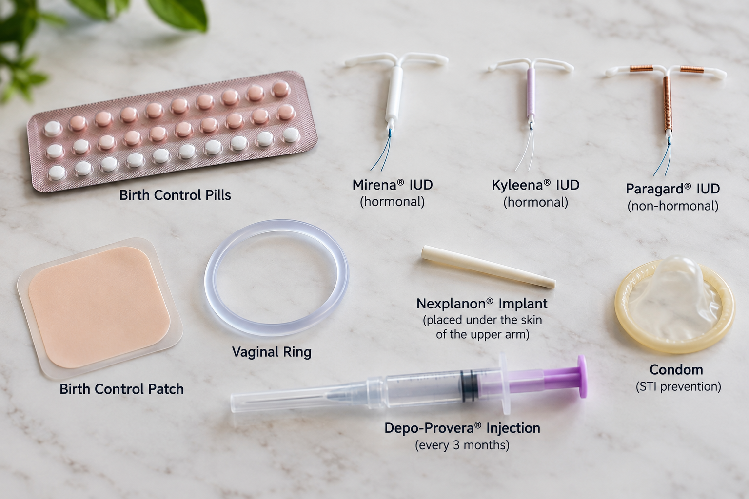
Common contraceptive methods available for pregnancy prevention.

Gynecologic Care
Preventive Care for Every Stage of a Woman's Life
Preventive gynecologic care is one of the most important investments you can make in your long-term health. Regular visits allow us to detect potential health concerns early, prevent disease whenever possible, and help you make informed decisions about your reproductive and overall wellness. At our practice, preventive care is personalized to your age, lifestyle, medical history, and future health goals. Whether you are a teenager visiting for your first gynecologic appointment, planning a pregnancy, managing menopause, or simply maintaining your health, we are committed to providing compassionate, evidence-based care. Our goal is not only to treat illness—but to help you stay healthy.
What Is Preventive Gynecologic Care?
Preventive care focuses on identifying health concerns before they become serious. During your annual well-woman examination, we evaluate your overall reproductive health, discuss any concerns you may have, review recommended screenings, and develop a plan tailored specifically to you. Even if you feel healthy and have no symptoms, annual visits remain essential because many gynecologic conditions develop silently before symptoms appear.
Annual Well-Woman Examination
- Your annual exam may include:
- Comprehensive medical and family history review
- Blood pressure measurement
- Height, weight, and BMI evaluation
- Breast examination
- Pelvic examination when appropriate
- Cervical cancer screening (Pap test and HPV testing as recommended)
- Discussion of menstrual concerns
- Birth control counseling
- Preconception counseling
- Menopause management
- Sexual health evaluation
- Screening for sexually transmitted infections (STIs) when indicated
- Review of medications and supplements
- Lifestyle counseling including nutrition, exercise, and weight management
Every visit is individualized based on your age, symptoms, risk factors, and current medical guidelines.
Cervical Cancer Screening
Routine cervical cancer screening has dramatically reduced deaths from cervical cancer through early detection of abnormal cells before cancer develops. Depending on your age and medical history, screening may include:
Pap test Human Papillomavirus (HPV) testing Co-testing (Pap test with HPV testing) Most abnormal Pap tests do not mean cancer. Detecting abnormal cells early allows treatment before they progress.
HPV Prevention
Human Papillomavirus (HPV) is the most common sexually transmitted infection and is responsible for nearly all cervical cancers.
- Preventive care includes
- HPV vaccination recommendations
- Routine cervical cancer screening
- Education regarding HPV transmission
- Follow-up for abnormal cervical testing
Vaccination is most effective before exposure to HPV but remains beneficial for many adults even after exposure.
Breast Health
Routine breast health evaluations remain an important part of preventive care.
- Your visit may include
- Clinical breast examination
- Education regarding breast self-awareness
- Mammogram recommendations based on age and risk factors
- Evaluation of breast pain, lumps, nipple discharge, or skin changes
Women with significant family histories may benefit from earlier screening or genetic counseling.
Sexually Transmitted Infection (STI) Screening
Many sexually transmitted infections cause few or no symptoms but can lead to infertility, chronic pelvic pain, pregnancy complications, and increased cancer risk if left untreated.
- Screening may include testing for:
- Chlamydia
- Gonorrhea
- HIV
- Syphilis
- Hepatitis B
- Hepatitis C
- Trichomonas
Testing recommendations depend upon age, risk factors, pregnancy status, and sexual history, including contraceptive counseling and birth control choices.
- We offer counseling regarding:
- Birth control pills
- Hormonal IUDs
- Copper IUDs
- Nexplanon implant
- Depo-Provera injection
- Vaginal ring
- Contraceptive patch
- Permanent sterilization
- Natural family planning
Together, we can select the method that best fits your health, lifestyle, and reproductive goals.
Menstrual Health
Heavy, painful, irregular, or absent periods are not something you simply have to live with.
- Preventive visits provide an opportunity to evaluate:
Early diagnosis often leads to simpler and more effective treatment.
If you are planning pregnancy, a preconception visit can improve the chances of a healthy pregnancy.
- Topics include:
- Prenatal vitamins and folic acid
- Medication review
- Vaccination status
- Chronic disease manage ment
- Genetic carrier screening
- Weight optimization
- Lifestyle recommendations
- Fertility planning
Healthy pregnancies begin before conception!
Menopause and Healthy Aging
Preventive care continues well beyond the reproductive years.
- We provide guidance for:
- Hot flashes
- Night sweats
- Vaginal dryness
- Hormone replacement therapy
- Bone health
- Osteoporosis prevention
- Cardiovascular risk reduction
- Urinary symptoms
- Sexual wellness after menopause
Our goal is to help women remain healthy and active throughout every stage of life.
Bone Health
Women experience accelerated bone loss after menopause.
- Preventive recomendations may include:
- Bone Health
- Calcium intake
- Vitamin D supplementation
- Weight-bearing exercise
- Fall prevention
- Bone density testing (DEXA scan) when appropriate
Early intervention can significantly reduce fracture risk.
Heart Health
Heart disease remains the leading cause of death among women.
- Your annual visit may include discussion of:
- Blood pressure
- Weight management
- Cholesterol screening
- Diabetes risk
- Smoking cessation
- Exercise
- Healthy nutrition
- Family history of cardiovascular disease
Maintaining heart health is an essential part of women's preventive care.
Vaccinations
Preventive care also includes keeping vaccinations current.
- Depending on your age and health, recommended vaccines may include:
- HPV vaccine
- Influenza vaccine
- Tdap
- Shingles vaccine
- Pneumonia vaccine
- RSV vaccine for eligible patients
We can help determine which immunizations are appropriate for you.
Lifestyle and Wellness Counseling
Preventive medicine extends beyond medical testing.
- We provide counseling regarding:
- Healthy nutrition
- Weight management
- Exercise
- Sleep
- Stress reduction
- Mental health
- Smoking cessation
- Alcohol moderation
- Healthy relationships
- Sexual wellness
Small lifestyle changes often have a significant impact on long-term health. When Should You Schedule Your Annual Visit?
- You should schedule a preventive gynecologic examination:
- Every year, even if you feel healthy
- If you develop abnormal bleeding
- If you experience pelvic pain
- If you notice breast changes
- If you have concerns about birth control
- Before becoming pregnant
- During menopause
- Whenever you have questions regarding your reproductive health
Annual visits allow us to identify problems early and provide the most effective care.
Our Commitment to Women's Health
Preventive care is about more than annual exams—it's about building a lifelong partnership in your health. We are dedicated to providing comprehensive, compassionate care tailored to your individual needs. From adolescence through menopause and beyond, we are here to support every stage of your health journey.
Whether you need routine screenings, contraception counseling, menopause management, or guidance on maintaining a healthy lifestyle, our team is committed to helping you achieve your best health.
Schedule your annual well-woman examination today and take an important step toward protecting your health for years to come.
Frequently Asked Questions
How often should I have a well-woman exam?
Most women should have a preventive gynecologic visit every year. The frequency of Pap tests may differ based on your age and previous results, but annual wellness visits remain important
Do I need a Pap test every year?
Not necessarily. Current guidelines recommend Pap tests at specific intervals depending on your age and HPV status. Your provider will recommend the screening schedule that is right for you.
What if I don't have any symptoms?
Many gynecologic conditions—including cervical abnormalities, high blood pressure, and some sexually transmitted infections—may not cause symptoms in their early stages. Routine preventive care helps identify these conditions before they become more serious.
Can I discuss birth control or menopause during my annual visit?
Yes. Your preventive visit is an excellent opportunity to discuss contraception, menstrual concerns, menopause symptoms, fertility planning, sexual health, and any other questions you may have.

What is the purpose of a Pap test?
A Pap test, also called a Pap smear or Papanicolaou test, checks for abnormal cells on the cervix which is the lower part of the uterus that opens into the vagina. Sometimes these changes are precancerous, meaning they could develop into cervical cancer if left untreated. Most cervical cancers develop gradually over many years. Regular screening, appropriate follow-up, and treatment of precancerous changes can prevent most cases of cervical cancer.
How is a Pap test performed?
While you lie on an examination table, your healthcare provider gently inserts a speculum into the vagina to see the cervix. A small brush or spatula is used to collect cells from the surface of the cervix. These cells are sent to a laboratory, where they are examined for abnormalities.
Collecting the sample takes only a few moments. You may feel mild pressure or brief discomfort. Let your provider know if you experience pain so adjustments can be made.
What is the purpose of testing for HPV?
Human papillomavirus (HPV) is a common virus that causes nearly all cervical cancers. A Pap test looks for abnormal cells, while an HPV test looks for the high-risk types of HPV that can cause those changes. HPV testing helps identify women who may need closer monitoring, sometimes before abnormal cells appear.
A positive HPV test does not mean you have cancer or will develop cancer. Most HPV infections clear on their own. When a high-risk infection persists, however, it can cause precancerous changes over time. HPV types 16 and 18 cause most cervical cancers; type 45 is another important high-risk type.
The Gardasil 9 vaccine protects against seven high-risk HPV types, including 16, 18, and 45, as well as two types that cause most genital warts. Vaccination helps prevent new HPV infections but does not treat an existing infection. Regular cervical cancer screening remains important even after vaccination. Ask your pediatrican about vaccinating your child. If you are an adult, you can still get the vaccine.

From left to right, the above diagram displays the single layer columnar cells next to the opening of the center of the cervix, to the squamocolumnar junction where cells change from columnar to squamous cells.
The opening of the cervix, the cervical os, is surrounded by columnar cells which are one layer bright pink cells. These cells undergo metaplasia to develop into multiple layers of flat or squamous cells. When these cells change form they can become damaged resulting in abnormal cells that can lead to cervical cancer.
What is Cervical Metaplasia?
Cervical metaplasia is a normal process in which the glandular cells lining the inner cervix are replaced by squamous cells, which are better suited to the acidic environment of the vagina. This change occurs in an area called the transformation zone or squamocolumnar junction as seen on the diagram, where the inner and outer portions of the cervix meet.
Although metaplasia itself is not abnormal or precancerous, the immature cells in the transformation zone are particularly susceptible to infection with high-risk types of human papillomavirus (HPV).Metaplasia is at its greatest in puberty. This is why sexual intercourse at a very young age increase the risk of cervical cancer.
Metaplasia is a normal change in cell type, whereas dysplasia represents abnormal cellular growth and maturation. Metaplasia does not inevitably progress to dysplasia; persistent high-risk HPV infection is the critical factor.
Cervical Intraepithelial Neoplasia (CIN)
- CIN is defined as separate grades based on the depth of change through the layers of the squamous cervical cells.
- CIN I: Mild changes involving approximately the lower one-third of the lining. Often resolves on its own; it is not cancer.
- CIN II: Moderate changes involving approximately the lower two-thirds. A precancerous change that may regress or progress.
- CIN III: Severe changes involving more than two-thirds, sometimes the full thickness. A significant precancerous change, but not invasive cancer.
- CIS (Carcinoma In Situ An older term for full-thickness abnormal squamous cells that remain confined to the lining. Included within CIN 3, rather than a separate stage after it.
- Invasive Cervical Cancer: Cancer cells have crossed the basement membrane—the boundary beneath the lining—and entered the underlying cervical tissue. CIN and CIS remain within the lining; invasion through the basement membrane is the key distinction.
ASC-US, LSIL, HSIL, AIG, and AIS
- ASC-US: Atypical Cells of Undetermined Significance. The squamous cells collected during a Pap test look slightly abnormal, but the changes are not clear enough to classify as LSIL or HSIL. This can occur due to inflamation, irritation or low estrogen such as in menopause. IF HPV is present, it may be the cells will later become LSIL or HSIL.
- LSIL and HSIL describe Pap test findings. CIN I, II, III, and CIS describe biopsy findings.
- LSIL: Low-Grade Squamous Intraepithelial lesions, generally is the same as CIN I.
- HSIL: High-Grade Squamous Intraepithelial lesions include CIN II, CIN III and CIS because the are all treated the same.
- AGC: Atypical Glandular Cells are abnormal cervical glandular cells which may or may not be a precursor to cancer.
- AIS: Adenocarcinoma In Situ is different from CIN as it involves glandular cells rather than squamous cells and remains a current, distinct diagnosis.
Risk factors for cervical cancer
The risk factors for squamous cell and glandular cell cervical cancer are essentially the same.
- Persistent high-risk HPV, especially with HPV-16 and HPV-18
- Weakened immune system
- Smoking
- Multiple sexual partners
- Sexual activity at a young age
- Long-term oral contraceptive use
- Having several full-term pregnancies
- Inadequate cervical screening
How are Abnormal Pap Tests Treated?
When a Pap test reveals abnormal cells, the next step may be a procedure called colposcopy. A colposcope is a specialized, low-power microscope that allows your gynecologist to examine the cervix in greater detail. During the examination, a mild vinegar solution (acetic acid) is applied to the cervix, causing certain abnormal areas to temporarily turn white and become easier to identify. If a suspicious area is found, a small tissue sample (biopsy) may be taken and sent to a pathology laboratory for examination. The biopsy helps determine whether precancerous changes are present and whether further treatment is necessary.
Treatment for ASC-US
When ASC-US is found, typically an HPV test will be peformed as well. If the HPV is negative a colposcopy is not necessary. You may be asked to repeat your Pap Test in six to twelve months. If high risk HPV is found, a colposcopy with biopsy may be necessary. If HPV was not performed you may be asked to repeat your Pap with HPV testing in the near future.
Treatment for LSIL and HSIL
A colposcopy is usually done with a finding do LSIL. A biopsy of any abnormal appearing tissue is done and sent to pathology.
Treatment for AGC
A colposcopy is peformed along with endocervical sampling and possibly an endometrial biopsy. The next step depends on the results.
Treatment for AIS
AIS is already a confirmed precancerous glandular lesion, not just a screening abnormality. Therefore, a colposcopy is not necessary. The next step is usually a cone biopsy to exclude invasive adenocarcinoma and to determine the status of the margins of the biopsy.
Possible Cervical Biopsy Results
- Normal cervical tissue: No abnormal or precancerous cells are identified.
- Chronic cervicitis. Inflammation of the cervix: which may be associated with irritation, infection, or other benign conditions.
- Squamous metaplasia:normal, benign process in which glandular cells are replaced by squamous cells, commonly occurring in the transformation zone.
- HPV-related changes (koilocytosis): Cellular changes associated with human papillomavirus infection, sometimes without sufficient abnormalities to diagnose CIN 1.
- CIN I (mild dysplasia): Abnormal cells generally involving the lower third of the cervical epithelium. Usually associated with transient HPV infection and frequently resolves without treatment.
- CIN II (moderate dysplasia): More significant abnormalities, historically described as involving approximately the lower two-thirds of the epithelium. Considered a high-grade precancerous lesion.
- CIN III (severe dysplasia): Markedly abnormal cells involving most or all of the epithelial thickness. A high-grade precancerous lesion with greater potential to progress to invasive cancer if untreated.
- Carcinoma in situ (CIS): An older term generally corresponding to full-thickness CIN 3 without invasion through the basement membrane.
- Adenocarcinoma in situ (AIS) A precancerous lesion involving the glandular cells of the cervix rather than the squamous epithelium.
Biopsies that Indicate Cancer
- Invasive squamous cell carcinoma: Cancer arising from squamous cells that has invaded beyond the epithelial basement membrane.
- Invasive adenocarcinoma Cancer arising from glandular cells of the cervix that has invaded the underlying tissue.
The Next Step in Treatment
Your treatment and follow-up depend on the biopsy result, your previous Pap and HPV results, your age, and your pregnancy plans. A normal or mildly abnormal biopsy may require additional evaluation if the Pap suggested a higher-grade abnormality.
-
Normal cervical tissue:
No precancerous or cancerous changes were found.
Next step: No treatment is needed for the tissue itself. After a low-grade screening abnormality, repeat HPV testing, with or without a Pap test, is often recommended in one year. Earlier or additional evaluation may be needed based on your previous results. -
Chronic cervicitis:
Inflammation of the cervix, which may be associated with irritation,
infection, or other benign conditions.
Next step: Often no treatment is needed if there are no symptoms or signs of infection. If an infection is identified or suspected, appropriate treatment is prescribed. Inflammation on a biopsy alone does not necessarily require antibiotics. -
Squamous metaplasia:
A normal, benign process in which glandular cells are replaced by
squamous cells, commonly occurring in the transformation zone.
Next step: No treatment is needed. Follow-up testing is based on your Pap and HPV results and screening history. -
HPV-related changes (koilocytosis):
Cellular changes associated with human papillomavirus infection,
sometimes without enough abnormalities to diagnose CIN 1.
Next step: Monitoring is usually recommended, often with repeat HPV testing, with or without a Pap, in one year after a low-grade screening result. There is no medication that eliminates cervical HPV itself. -
CIN I (mild dysplasia):
Abnormal cells generally involving the lower third of the cervical
lining. These changes often resolve without treatment.
Next step: Observation is preferred, usually with repeat testing in one year after a low-grade screening result. Treatment may be considered if CIN 1 persists for at least two years. -
CIN II (moderate dysplasia):
A high-grade precancerous change, historically described as
involving approximately the lower two-thirds of the cervical lining.
Next step: Treatment to remove the abnormal tissue, usually with a LEEP or cone biopsy, is often recommended. Carefully selected patients, particularly younger patients or those concerned about future pregnancy, may instead undergo close monitoring at six-month intervals. -
CIN III (severe dysplasia):
A high-grade precancerous change involving most or all of the
thickness of the cervical lining.
Next step: Treatment is recommended, usually with a LEEP or cone biopsy to remove the abnormal tissue and evaluate it for hidden cancer. Continued follow-up is needed after treatment. -
Carcinoma in situ (CIS):
An older term generally corresponding to full-thickness CIN 3,
without invasion into the underlying tissue. This is not invasive cancer.
Next step: Treatment is the same as for CIN 3, usually with a LEEP or cone biopsy, followed by ongoing surveillance. -
Adenocarcinoma in situ (AIS):
A precancerous change involving the glandular cells of the cervix.
Next step: An excisional procedure, often a cone biopsy, is needed to evaluate for invasive cancer and determine whether the abnormal tissue has been completely removed. Hysterectomy is generally recommended when childbearing is complete. Selected patients wishing to preserve fertility may be managed with excision and close follow-up if the margins are clear.
Biopsies that Indicate Cancer
-
Invasive squamous cell carcinoma:
Cancer arising from squamous cells that has invaded the underlying tissue.
Next step: Prompt referral to a gynecologic oncologist is recommended to determine the stage and treatment plan. Treatment may include surgery, radiation, chemotherapy, or a combination, depending on the extent of the cancer. -
Invasive adenocarcinoma:
Cancer arising from glandular cells of the cervix that has invaded
the underlying tissue.
Next step: Prompt referral to a gynecologic oncologist is recommended for staging and treatment planning. Treatment depends on the stage and may include surgery, radiation, chemotherapy, or other cancer therapies.
When is a Referral to a Gynecologic Oncologist Necessary?
Most abnormal Pap tests do not indicate cancer. Even when a cervical biopsy shows precancerous changes, these can usually be treated by your regular gynecologist. However, if a biopsy reveals cervical cancer, or if there is a strong suspicion of invasive cancer, you will be referred to a gynecologic oncologist, a physician who specializes in diagnosing and treating cancers of the female reproductive system.
There are two main types of cervical cancer:
- Squamous Cell Carcinoma: This is the most common type of cervical cancer, accounting for approximately 70–80% of cases. It develops from the squamous cells covering the outer portion of the cervix and is usually associated with persistent infection with high-risk human papillomavirus (HPV).
- Adenocarcinoma: This type accounts for approximately 20–25% of cervical cancers and develops from the glandular cells lining the cervical canal. Because these cells are located higher inside the cervix, adenocarcinoma can sometimes be more difficult to detect during routine screening. Most cervical adenocarcinomas are associated with high-risk HPV, although some less common types are not.
Adenocarcinoma in situ (AIS) is a precancerous condition involving the glandular cells of the cervix. Although it is not invasive cancer, it requires careful evaluation and treatment because it can coexist with an invasive cancer or progress to cancer if left untreated. Referral to a gynecologic oncologist may be recommended, particularly when the diagnosis is uncertain or more extensive treatment is being considered.
What Should I Expect if Cervical Cancer Is Diagnosed?
Your gynecologic oncologist will review your biopsy results, perform a pelvic examination, and determine whether additional imaging or testing is necessary to establish the extent, or stage, of the cancer. Treatment depends on the type and stage of cancer, your overall health, and whether you wish to preserve fertility.
Very early cervical cancers may be treated with a cone biopsy or surgery. More advanced cancers may require a combination of radiation therapy, chemotherapy, immunotherapy, or other specialized treatments. Some patients with early-stage disease may be candidates for fertility-preserving treatment.
Most importantly, an abnormal Pap test does not mean you have cervical cancer. Cervical cancer usually develops slowly, and screening allows us to identify and treat precancerous changes long before invasive cancer develops. Even when cancer is diagnosed, early detection greatly improves the likelihood of successful treatment.
```During pregnancy, precancerous changes are generally monitored, with treatment deferred until after delivery unless cancer is suspected.

Polyendocrine Metabolic Ovarian Syndrome (PMOS / PCOS)
In May 2026, an international consensus of medical professionals, researchers, and patients established the new name, polyendocrine metabolic syndrome, to better reflect the complex nature of this condition. The older term, polycystic ovarian syndrome (PCOS), was misleading because it suggested that ovarian cysts were the primary problem. In reality, PMOS involves multiple hormonal and metabolic abnormalities that can affect menstrual cycles, fertility, insulin function, cardiovascular health, and overall well-being.
Understanding PMOS
Polyendocrine metabolic ovarian syndrome (PMOS) is one of the most common hormonal and metabolic disorders affecting women of reproductive age.
Although women with PMOS may have multiple small follicles visible on ovarian ultrasound, these are not true ovarian cysts. In fact, some women with PMOS have completely normal-appearing ovaries on ultrasound. The condition involves a complex interaction of reproductive hormones, androgen production, and metabolic function, including insulin resistance in many patients.
Many women first seek medical attention because of irregular menstrual periods, difficulty becoming pregnant, acne, or unwanted facial and body hair (hirsutism). Others are concerned about weight gain or difficulty losing weight. However, PMOS can also occur in women who are not overweight.
Because PMOS can affect long-term health as well as reproductive function, evaluation should extend beyond the ovaries to include metabolic and cardiovascular risk factors.
How Is PMOS Diagnosed?
In adults, PMOS is generally diagnosed when at least two of the following are present, after other causes have been excluded:
- Irregular or infrequent ovulation: often reflected by irregular or absent menstrual periods.
- Excess androgen activity: such as increased facial hair, or elevated testosterone on blood testing.
- Polycystic ovarian morphology: identified by ultrasound, or in some adults by an appropriately measured anti-Müllerian hormone (AMH) level.
When irregular cycles and excess androgen activity are both present, an ultrasound is not necessarily needed to establish the diagnosis. Different criteria apply to adolescents; ultrasound and AMH are not recommended to diagnose PMOS during adolescence.
Why PMOS Matters Beyond Menstrual Periods
PMOS is associated with an increased risk of insulin resistance, prediabetes, type 2 diabetes, abnormal cholesterol levels, high blood pressure, and sleep apnea. Cardiovascular risk factors deserve attention, although having PMOS does not mean that heart disease is inevitable.
Women who ovulate infrequently may go months without a menstrual period. Prolonged exposure of the uterine lining to estrogen without regular progesterone exposure increases the risk of endometrial hyperplasia and endometrial cancer. Fortunately, there are effective ways to protect the uterine lining.
Pregnancy with PMOS can carry higher risks of gestational diabetes, hypertensive disorders, and certain other complications. These risks can often be addressed through appropriate preconception care and prenatal monitoring. Fertility medications require individualized monitoring; the risks of multiple pregnancy and ovarian hyperstimulation vary considerably by treatment.
What Causes PMOS?
There is no single known cause. PMOS involves a combination of genetic susceptibility, altered ovarian hormone production, and, in many patients, insulin resistance. Higher insulin levels can stimulate ovarian androgen production and reduce sex hormone-binding globulin (SHBG), increasing the amount of biologically active testosterone.
Common findings include unwanted coarse facial or body hair (hirsutism), acne, scalp hair thinning, and sometimes acanthosis nigricans—dark, velvety skin often seen on the neck or in skin folds. Not everyone with PMOS has these findings, and obesity is not required for diagnosis.
Laboratory Tests: What We Check and Why
There is no single blood test that confirms PMOS. I select laboratory studies based on your symptoms, menstrual history, medications, and examination. Some tests help document excess androgens, others exclude conditions that resemble PMOS, and others look for metabolic complications.
Hormone and Diagnostic Tests
- Total and free testosterone: assess androgen excess that may contribute to hirsutism, acne, and irregular ovulation. Reliable laboratory methods are important. Hormonal birth control can affect results.
- DHEA-S: may help assess adrenal androgen production, particularly when testosterone results do not explain symptoms or androgen excess is pronounced.
- 17-hydroxyprogesterone: screens for nonclassic congenital adrenal hyperplasia, a hormonal disorder that can resemble PMOS; often obtained in the morning.
- TSH: checks for thyroid disease, which can cause irregular periods and other overlapping symptoms.
- Prolactin: checks for elevated prolactin, another possible cause of absent or irregular menstrual periods.
- Pregnancy test: appropriate when a menstrual period is missed and pregnancy is possible.
- FSH and estradiol: may be useful in selected patients with absent periods to evaluate ovarian function or other causes; they are not required routinely for every patient.
- AMH: may help assess polycystic ovarian morphology in adults in appropriate circumstances, but is not a stand-alone diagnostic test and should not routinely be ordered in addition to ultrasound for the same purpose.
What about the LH/FSH ratio? Although an elevated ratio was historically associated with PMOS, it is not sufficiently reliable to diagnose or exclude the condition. Likewise, routine fasting insulin measurements are not recommended because available assays and cutoffs do not reliably guide individual care.
Metabolic and Other Tests
- 75-gram, two-hour oral glucose tolerance test (OGTT): the most accurate recommended test for detecting impaired glucose tolerance and type 2 diabetes in women with PMOS, regardless of BMI. It is especially useful when planning pregnancy or fertility treatment.
- Hemoglobin A1c or fasting glucose: alternatives when an OGTT cannot be performed, although they may miss some abnormalities.
- Lipid panel: measures cholesterol and triglycerides to assess cardiovascular risk.
- Blood pressure: checked during office visits because hypertension is more common with PMOS.
- Additional tests when indicated: a complete blood count and ferritin for heavy bleeding or suspected iron deficiency; liver tests or other studies when suggested by metabolic risk, symptoms, or treatment plans.
Ultrasound may help evaluate ovarian appearance or investigate abnormal uterine bleeding, but it is not necessary for every patient. If bleeding is prolonged or unusually heavy, an endometrial evaluation may also be appropriate depending on age and risk factors.
Treatment: A Personalized Approach
My goal is not simply to make menstrual periods regular. It is to address metabolic health, protection of the uterine lining, bothersome androgen-related symptoms, and fertility goals. Treatment depends on what matters most to each patient.
Nutrition, Physical Activity, and Weight Management
Regular physical activity and good nutrition improve metabolic health, even when weight does not change. Both aerobic activity and resistance training can help improve insulin sensitivity. There is no single diet or exercise program proven to be best for every woman with PMOS.
For women who have excess weight, even modest weight loss—sometimes around 5% of starting weight—may improve menstrual regularity and metabolic measures. For example, 5% of 240 pounds is 12 pounds. However, women of every body size can benefit from healthy habits, and not all women with PMOS need to lose weight.
Exercise plans should be adapted to fitness, health conditions, and personal preference. A routine cardiac stress test is not required simply because a previously inactive person is over age 35; symptoms and cardiovascular risk determine whether additional evaluation is needed.
Hormonal Treatment When Pregnancy Is Not Desired
Combined hormonal contraceptives are often first-line treatment for irregular periods, acne, and unwanted hair growth when pregnancy is not desired and estrogen is medically appropriate. They suppress ovarian androgen production and increase SHBG. Selection should consider blood pressure, migraine history, smoking, blood clot risk, and other individual factors.
Some formulations have less androgenic or antiandrogenic progestins, but there is no single birth control pill that is best for everyone. I do not consider a particular brand automatically “wrong” for every patient with PMOS.
If estrogen-containing contraception is unsuitable, cyclic progestin treatment or a levonorgestrel-releasing IUD may be used to protect the uterine lining. These options do not necessarily treat acne or excess hair growth.
Metformin and Metabolic Treatment
Metformin improves insulin sensitivity and can be especially useful when PMOS is accompanied by metabolic risk factors or impaired glucose regulation. It may also improve menstrual regularity in some patients, but it is not a substitute for all other treatments and is not generally the most effective first-line medication for unwanted facial hair.
For selected patients, additional weight-management medications may be considered under appropriate medical supervision, with careful attention to pregnancy plans and medication safety.
Unwanted Facial Hair and Acne
Hormonal contraceptives may help reduce androgen-related symptoms. When needed, medications such as spironolactone can be considered for hirsutism, usually alongside reliable contraception because antiandrogen medications can harm a developing male fetus. Laser hair removal, electrolysis, and dermatologic treatments may also be helpful.
When Pregnancy Is Desired
Many women with PMOS conceive naturally, while others need assistance with ovulation. For anovulatory infertility related to PMOS, letrozole is generally the preferred first-line ovulation-induction medication when no other infertility factors are present. An infertility evaluation may also include semen analysis and assessment of the fallopian tubes when appropriate.
Optimizing glucose control, blood pressure, nutrition, and other health conditions before conception can improve pregnancy planning. Fertility treatment should be individualized and monitored to reduce avoidable complications.
Long-Term Follow-Up
PMOS is a long-term condition, but its symptoms and health risks can often be managed successfully. Follow-up may include monitoring menstrual patterns, blood pressure, cholesterol, and glucose status. Glucose testing is generally repeated every one to three years according to individual risk factors, or sooner when clinically indicated.
My approach is to treat the whole patient—not just the ovaries. By identifying hormonal and metabolic concerns early, we can develop a treatment plan that supports your current symptoms, future fertility, and long-term health.
Reference:
Teede HJ, Bahri Khomami M, Morman R, et al.
Polyendocrine metabolic ovarian syndrome,
the new name for polycystic ovarian syndrome:
a multistep global consensus process.
The Lancet. 2026.
Read article

Uterine Fibroids
Uterine fibroids are extremely common, and I see women with fibroids in my practice almost every day. Fibroids, also called leiomyomas, are benign (noncancerous) growths that develop from the muscular wall of the uterus. Although they can occur at any age during the reproductive years, they become increasingly common after age 30.
Many women have fibroids without ever knowing it. Others experience heavy menstrual bleeding, pelvic pressure, discomfort, or occasionally problems with fertility. Fortunately, most fibroids do not require treatment.
If you have been diagnosed with fibroids but are not experiencing any symptoms, there is usually no reason to remove them. In many cases, simply monitoring the fibroids is the best approach.
What Symptoms Can Fibroids Cause?
The most common symptom associated with uterine fibroids is heavy or prolonged menstrual bleeding. Menstrual periods lasting longer than seven days, frequent passage of large blood clots, or bleeding that interferes with normal daily activities should be evaluated.
Over time, excessive menstrual bleeding can lead to iron deficiency and anemia, sometimes severe enough to require medical or surgical treatment.
Although fibroids do not always cause pain, larger fibroids can produce pressure on surrounding pelvic organs. Depending on their size and location, symptoms may include:
- Heavy or prolonged menstrual periods
- Pelvic pressure or discomfort
- Frequent urination or difficulty emptying the bladder
- Constipation or rectal pressure
- Pain during sexual intercourse (dyspareunia)
- Abdominal enlargement or a sensation of fullness
- Occasionally, infertility or pregnancy complications
The severity of symptoms does not always correspond to the size of the fibroid. A relatively small fibroid located inside the uterine cavity may cause significant bleeding, while a much larger fibroid on the outside of the uterus may cause few problems.
Different Types of Fibroids
Fibroids are classified according to their location within or around the uterus. Their location is often more important than their size when determining whether treatment is necessary.
Subserosal Fibroids
These develop beneath the outer surface of the uterus and grow outward into the pelvis. They may become quite large without causing significant menstrual bleeding. However, they can create pressure on the bladder, bowel, or other pelvic structures.
Intramural Fibroids
These are the most common type of fibroid and develop within the muscular wall of the uterus. Many cause no symptoms, but larger intramural fibroids can enlarge or distort the uterus and contribute to heavy menstrual bleeding or pelvic pressure.
Submucosal Fibroids
These grow immediately beneath the endometrium, the lining of the uterus, and may protrude into the uterine cavity. Submucosal fibroids are particularly likely to cause heavy or irregular bleeding, even when they are relatively small. They may also interfere with fertility or increase the risk of miscarriage.
Pedunculated Fibroids
These fibroids are attached to the uterus by a narrow stalk. They may grow outward from the uterus or project into the uterine cavity. Occasionally, a pedunculated fibroid can twist on its stalk, producing sudden and sometimes severe pain.
Intracavitary Fibroids
These project substantially or entirely into the uterine cavity, sometimes attached by a stalk. They frequently cause abnormal bleeding and may be particularly suitable for removal through a hysteroscope without an abdominal incision.
When Should Fibroids Be Treated?
The decision to treat fibroids depends on several factors, including the severity of symptoms, the size and location of the fibroids, your age, and whether you wish to become pregnant in the future.
Treatment is generally considered when fibroids cause significant bleeding, anemia, persistent pain, pressure on surrounding organs, or certain fertility problems.
There are several effective treatment options, ranging from medications and minimally invasive procedures to surgery. Not every treatment is appropriate for every woman.
Hysterectomy: The Definitive Treatment
A hysterectomy, or surgical removal of the uterus, is the only treatment that permanently eliminates uterine fibroids and prevents new ones from developing.
Depending on the size of the uterus, previous surgeries, and other individual factors, a hysterectomy may be performed through the vagina, laparoscopically, robotically, or through a traditional abdominal incision.
One important point I emphasize to my patients is that removing the uterus does not necessarily mean removing the ovaries.
The ovaries produce hormones that contribute to bone health, cardiovascular health, and other important functions. In women who have not reached menopause, preserving healthy ovaries is often preferable unless there is a medical reason to remove them. Even after menopause, the ovaries continue to produce small amounts of hormones.
For women who have completed childbearing and are experiencing significant symptoms from fibroids, hysterectomy may be an excellent option.
Endometrial Ablation
Endometrial ablation is a minimally invasive procedure designed to reduce heavy menstrual bleeding by destroying the endometrium, the lining of the uterus that is shed during menstruation.
Unlike hysterectomy or myomectomy, endometrial ablation does not remove fibroids. It treats the bleeding rather than the fibroids themselves.
Ablation may be appropriate for selected women who have completed childbearing, have heavy menstrual bleeding, and have a uterine cavity that is not significantly distorted by fibroids.
Several methods of endometrial ablation have been developed over the years.
Rollerball Ablation
This is an older technique in which a hysteroscope and an electrical electrode are used to destroy the uterine lining. I performed many of these procedures earlier in my career, but newer technologies have largely replaced this approach.
Thermal Balloon Ablation
A small balloon is inserted into the uterine cavity and filled with heated fluid. The heat destroys the endometrial lining, reducing menstrual bleeding. This technique is now used less frequently.
NovaSure Endometrial Ablation
NovaSure uses radiofrequency energy to destroy the endometrial lining. A specially designed device is inserted through the cervix and expanded within the uterine cavity.
The treatment itself is generally brief and can be very effective for appropriately selected patients. However, fibroids that distort the uterine cavity may make this procedure less suitable.
Cryoablation
Cryoablation uses extremely cold temperatures to destroy the uterine lining. Certain systems can be performed in an office setting.
I previously performed office cryoablation but discontinued it because I was not as satisfied with the results compared with other approaches.
What Can You Expect After Ablation?
Most women recover relatively quickly, often returning to normal activities within a few days.
Some women stop menstruating altogether, while many continue to have periods that are considerably lighter. Others may experience a return of heavy bleeding months or years later and eventually require additional treatment.
Ablation is not a form of contraception, and pregnancy afterward can be dangerous. Women who undergo ablation should continue to use reliable contraception until menopause.
Another important consideration is that scarring after ablation may make future evaluation of the uterine lining more difficult. Abnormal bleeding, particularly after menopause, must still be investigated because it can be a warning sign of endometrial cancer.
For these reasons, ablation is not appropriate for every woman with heavy menstrual bleeding.
Uterine Artery Embolization (UAE)
Uterine artery embolization, sometimes called uterine fibroid embolization, is a minimally invasive procedure performed by an interventional radiologist.
During this procedure, a small catheter is introduced into an artery, usually through the wrist or groin, and guided into the arteries supplying the uterus. Tiny particles are then injected to reduce blood flow to the fibroids.
Without their normal blood supply, the fibroids gradually shrink, often improving heavy menstrual bleeding and pelvic pressure.
UAE can be effective for women with multiple fibroids as well as those with larger individual fibroids.
Although UAE allows a woman to keep her uterus, it may not be the best choice for women who wish to become pregnant. The effects on future fertility and pregnancy should be discussed carefully before proceeding.
Myomectomy: Removing Fibroids While Preserving the Uterus
A myomectomy is a surgical procedure in which fibroids are removed while the uterus is preserved.
Depending on the size, number, and location of the fibroids, myomectomy may be performed through:
- Hysteroscopy: A small instrument is passed through the cervix into the uterine cavity to remove selected submucosal fibroids.
- Laparoscopy or robotic surgery: Fibroids are removed through small abdominal incisions.
- Abdominal surgery: A larger incision is used when the fibroids are particularly large, numerous, or difficult to remove through minimally invasive techniques.
Myomectomy is often the preferred surgical treatment for women who wish to preserve their fertility, particularly when fibroids distort the uterine cavity.
However, removing fibroids does not prevent new ones from developing. Some women eventually require additional treatment.
Depending on the depth and location of the uterine incisions, a cesarean delivery may be recommended in future pregnancies.
Medications for Uterine Fibroids
Medications can sometimes control fibroid-related symptoms or temporarily reduce fibroid size, allowing women to avoid or postpone surgery.
Hormonal Contraceptives and Progestins
Birth control pills and certain other hormonal treatments may reduce menstrual bleeding but generally do not significantly shrink fibroids.
A levonorgestrel-releasing intrauterine device (IUD) can also be very effective in controlling bleeding when the uterine cavity is not substantially distorted.
Tranexamic Acid
Tranexamic acid is a nonhormonal medication taken during menstruation to reduce heavy bleeding. It does not shrink fibroids but may provide meaningful relief for women whose primary symptom is excessive menstrual flow.
GnRH Agonists — Lupron
Lupron (leuprolide acetate) is a medication that temporarily suppresses ovarian estrogen production, producing a reversible menopause-like state.
Because fibroids are sensitive to estrogen, reducing estrogen levels can cause them to shrink significantly.
I occasionally use Lupron for a short period before surgery, particularly when a woman has very large fibroids or significant anemia. Shrinking the fibroids and reducing menstrual bleeding beforehand may make surgery easier and safer.
Unfortunately, prolonged estrogen suppression can produce hot flashes, vaginal dryness, and loss of bone density. Fibroids also tend to grow again after treatment is discontinued.
For these reasons, Lupron is generally used for a limited period, sometimes with additional hormonal treatment to reduce side effects.
Newer GnRH Antagonists
Newer oral medications, including combinations containing relugolix or elagolix, can reduce heavy menstrual bleeding associated with fibroids.
These medications suppress hormonal stimulation of the fibroids and are generally prescribed with low doses of estrogen and progestin to reduce menopausal symptoms and protect bone density.
They can be effective alternatives for selected women who want to manage their symptoms without immediate surgery, although treatment duration, side effects, cost, and individual medical risks must be considered.
What Happens to Fibroids After Menopause?
Fibroids are influenced by reproductive hormones, particularly estrogen and progesterone. As hormone levels decline after menopause, fibroids commonly become smaller, and symptoms often improve.
For women approaching menopause who have relatively mild symptoms, observation or temporary medical treatment may be preferable to surgery.
However, new bleeding after menopause should never be assumed to be caused by fibroids. Postmenopausal bleeding requires evaluation to exclude other conditions, including endometrial cancer.
Choosing the Right Treatment
There is no single treatment that is best for every woman with uterine fibroids.
Some women need nothing more than periodic observation. Others benefit from medications or a minimally invasive procedure. For women with severe symptoms who have completed childbearing, hysterectomy may offer the most reliable and permanent solution.
My approach is to consider not simply the size of your fibroids, but how they are affecting your health, your daily activities, and your quality of life.
The goal is not necessarily to remove every fibroid. It is to choose the treatment that is safest, most effective, and most appropriate for you.

F
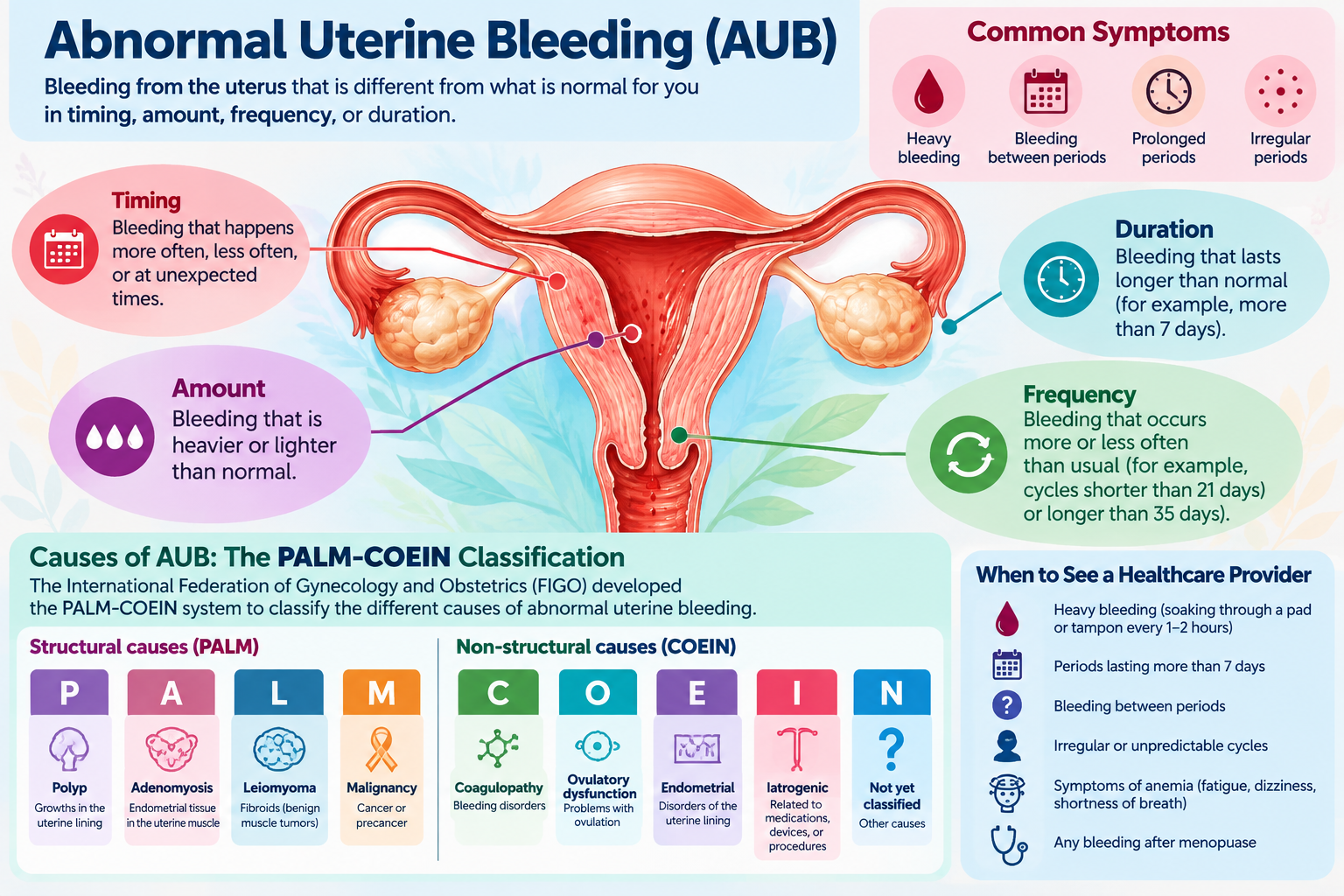
Understanding Abnormal Uterine Bleeding
Abnormal uterine bleeding (AUB) is one of the most common reasons women visit their gynecologist. Although menstrual periods vary from woman to woman, bleeding that is unusually heavy, occurs too frequently, lasts longer than expected, or happens between periods may be a sign of an underlying problem. Some women experience heavy bleeding with otherwise regular periods, while others may skip several months and then have prolonged or unpredictable bleeding. Bleeding after menopause is also abnormal and should always be evaluated.
Abnormal bleeding can interfere with daily activities, work, sleep, and overall quality of life. In some women, prolonged or heavy bleeding can lead to iron deficiency and anemia, causing fatigue, weakness, dizziness, or shortness of breath. The good news is that most causes of abnormal uterine bleeding can be successfully treated, often without surgery. The first step is identifying the cause.
What Causes Abnormal Uterine Bleeding?
Gynecologists use a classification system called by the acronym PALM-COEIN to describe the different causes of abnormal uterine bleeding. Although these letters may seem confusing, they simply help us organize the possible reasons for bleeding and select the most appropriate treatment. The first four categories (PALM) describe physical changes in the uterus that can often be identified with imaging or tissue examination. The remaining categories (COEIN) describe conditions related to hormones, blood clotting, medications, and other factors.
PALM
AUB-P: Polyps
Polyps are growths that develop from the lining of the uterus, called the endometrium. Most uterine polyps are benign (noncancerous), although a small percentage may contain precancerous or cancerous changes. Polyps may cause spotting between periods, heavier menstrual bleeding, or bleeding after menopause. Some women have no symptoms at all. Polyps can often be identified with ultrasound or a special ultrasound procedure called saline infusion sonography, in which a small amount of sterile fluid is placed inside the uterus to make the lining easier to examine. When treatment is necessary, polyps can usually be removed through a procedure called hysteroscopy, which allows the physician to look directly inside the uterus using a small camera.
AUB-A: Adenomyosis
Adenomyosis occurs when tissue similar to the lining of the uterus grows into the muscular wall of the uterus. This can cause the uterus to become enlarged and may lead to heavy, prolonged, or painful menstrual periods. Women with adenomyosis frequently experience severe menstrual cramps, pelvic pressure, or chronic pelvic discomfort. However, some women have relatively mild symptoms. Adenomyosis is more common in women in their later reproductive years but can occur at younger ages. Treatment may include hormonal medications, a progesterone-releasing intrauterine device (IUD), or other approaches to control bleeding and pain. Hysterectomy may be considered for women with severe symptoms who have completed childbearing and have not responded to less invasive treatment.
AUB-L: Leiomyomas (Uterine Fibroids)
Leiomyomas are commonly called uterine fibroids. These are noncancerous growths that develop from the muscle of the uterus. Fibroids are extremely common, particularly during the reproductive years. Many women have fibroids without ever developing symptoms. Whether a fibroid causes abnormal bleeding depends largely on its size and location. Fibroids that extend into or distort the uterine cavity are particularly likely to cause heavy or prolonged menstrual bleeding. Other symptoms may include pelvic pressure, frequent urination, constipation, and occasionally difficulty becoming pregnant. Treatment depends on the severity of symptoms, the size and location of the fibroids, the woman's age, and whether she wishes to preserve fertility. Options include medications, hormonal IUDs, procedures that remove or shrink fibroids, and, in selected cases, hysterectomy.
AUB-M: Malignancy and Endometrial Hyperplasia
This category includes precancerous changes and cancer involving the lining of the uterus. Endometrial hyperplasia occurs when the uterine lining becomes abnormally thick because of excessive growth of its cells. Some forms of hyperplasia carry a greater risk of developing into endometrial cancer. Women who experience prolonged exposure to estrogen without adequate progesterone, such as those with infrequent ovulation, may be at increased risk. Abnormal bleeding is often the first symptom of endometrial cancer. Fortunately, when detected early, endometrial cancer is frequently treatable. Evaluation may include ultrasound and an endometrial biopsy, in which a small sample of the uterine lining is obtained for examination. Any bleeding after menopause should be evaluated, even if it consists of only a small amount of spotting.
COEIN
AUB-C: Coagulopathy (Blood Clotting Disorders)
Some women experience heavy menstrual bleeding because their blood does not clot normally. One example is von Willebrand disease, an inherited bleeding disorder that can cause unusually heavy menstrual periods, frequent nosebleeds, easy bruising, or excessive bleeding following surgery or dental procedures. A bleeding disorder should be considered in women who have experienced very heavy periods since adolescence, particularly when there is a personal or family history of abnormal bleeding. Treatment depends on the underlying disorder and may involve medications that reduce menstrual blood loss or therapies directed at the clotting problem.
AUB-O: Ovulatory Dysfunction (Irregular Ovulation)
This is one of the most common causes of abnormal uterine bleeding. Normally, the ovaries release an egg approximately once each menstrual cycle. This process, called ovulation, helps regulate the production of estrogen and progesterone and produces predictable menstrual periods. When ovulation does not occur regularly, progesterone production may be inadequate. The uterine lining can continue growing under the influence of estrogen and eventually shed in an irregular or excessive manner. Women with ovulatory dysfunction may skip periods for weeks or months and then experience unusually heavy or prolonged bleeding.
- Polyendocrine Metabolic Syndrome (PMOS), aka PCOS/li>
- Adolescence, when menstrual cycles are still becoming established
- Perimenopause, when ovarian function becomes less predictable
- Thyroid Disorders
- Elevated prolactin levels
- Significant weight changes, excessive exercise, or physical stress
Common causes include:
AUB-E: Endometrial Causes
Sometimes a woman experiences heavy menstrual bleeding even though her uterus appears normal and she ovulates regularly. In these cases, the problem may involve the way the uterine lining controls bleeding during menstruation. Normally, the endometrium has mechanisms that limit blood loss and allow the lining to heal after each menstrual period. When these mechanisms do not function properly, menstrual bleeding may become excessive. Women with AUB-E typically have regular menstrual cycles but experience unusually heavy bleeding. Treatment may include nonsteroidal anti-inflammatory medications such as ibuprofen, tranexamic acid, hormonal contraceptives, or a progesterone-releasing IUD.
AUB-I: Iatrogenic Causes (Medications and Medical Treatments)
The word iatrogenic simply means that a condition is related to a medication or medical treatment. Certain medications and contraceptive methods can cause irregular or excessive uterine bleeding.
- Birth control pills, particularly when doses are missed
- Contraceptive implants
- Hormonal IUDs, especially during the first several months after insertion
- Injectable contraceptives
- Blood-thinning medications
- Certain hormone therapies
Examples include:
Bleeding associated with these treatments is not always dangerous, but persistent or troublesome bleeding should be evaluated rather than automatically attributed to the medication.
AUB-N: Not Otherwise Classified
This category includes less common causes of abnormal uterine bleeding that do not fit into the other groups. Examples include certain abnormalities of blood vessels within the uterus and defects in a previous cesarean section scar that can collect blood and cause spotting after menstrual periods. These conditions may require specialized imaging or additional evaluation to establish the diagnosis.
How Is Abnormal Uterine Bleeding Evaluated?
The evaluation begins with a careful review of your menstrual history, symptoms, medications, and overall health. Your physician may ask how often your periods occur, how long bleeding lasts, whether you pass large clots, and how frequently you need to change pads or tampons.
- Pregnancy testing: Pregnancy-related bleeding must be considered in women who could become pregnant.
- Complete blood count: Checks for anemia caused by excessive blood loss.
- Iron studies: May identify iron deficiency, even before significant anemia develops.
- Thyroid or other hormone testing: May be appropriate when hormonal abnormalities are suspected.
- Pelvic ultrasound: Helps identify fibroids, polyps, and other structural abnormalities.
- Endometrial biopsy: Samples the uterine lining to evaluate for precancerous or cancerous changes.
- Hysteroscopy:Allows direct examination of the uterine cavity when polyps, fibroids, or other abnormalities are suspected
- An endometrial biopsy is generally recommended for abnormal uterine bleeding in women age 45 and older. It may also be appropriate in younger women with certain risk factors, persistent bleeding, or bleeding that has not responded to treatment.
Depending on your age, symptoms, and medical history, testing may include:
Not every woman needs every test. The evaluation should be individualized.
Treatment Options for Abnormal Uterine Bleeding
- Treatment depends on the cause of the bleeding, the severity of symptoms, your age, your general health, and whether you wish to become pregnant in the future.
Many women can be treated successfully with medication.
- Nonhormonal treatments may include tranexamic acid, which helps reduce menstrual blood loss, or anti-inflammatory medications that can decrease both menstrual bleeding and cramps.
- Hormonal treatments may include birth control pills, progesterone therapy, or a levonorgestrel-releasing IUD. These treatments can help regulate bleeding, reduce the thickness of the uterine lining, and improve menstrual symptoms.
-
When a structural problem such as a polyp or fibroid is responsible, a procedure may be recommended.
- Hysteroscopy allows the physician to examine the uterine cavity and remove certain polyps or fibroids without an abdominal incision.
- Endometrial ablation is another option for selected women with heavy menstrual bleeding who have completed childbearing. It destroys or reduces the uterine lining to decrease bleeding. Pregnancy after ablation can be dangerous, so reliable contraception remains necessary.
- Hysterectomy provides definitive treatment for uterine bleeding but is generally reserved for women who have completed childbearing and have significant symptoms, certain serious diagnoses, or inadequate relief from other treatments.
When Should You Seek Medical Attention?
- if you experience unusually heavy periods, bleeding between periods, menstrual bleeding lasting longer than eight days, or unpredictable bleeding that interferes with your daily activities.
- Seek urgent medical care if you are soaking through a pad or tampon every hour for more than two consecutive hours, particularly if you feel dizzy, faint, weak, or short of breath. Severe bleeding, fainting, or significant abdominal pain may require emergency evaluation.
- Any bleeding during pregnancy requires appropriate assessment, and bleeding after menopause should always be discussed with your healthcare provider.
Contact your healthcare provider:
My Approach to Abnormal Uterine Bleeding
Every woman's menstrual history and health needs are different. My goal and my staff's goal is to identify the cause of abnormal bleeding, explain the available treatment options, and develop a plan that fits your individual circumstances.
For some women, reassurance and observation may be appropriate. Others may benefit from medication, office-based procedures, or surgery. Heavy or unpredictable menstrual bleeding should not be something you simply have to live with. With an accurate diagnosis and individualized treatment, most women can achieve significant improvement in their symptoms and quality of life.
References
- Munro MG, Critchley HOD, Fraser IS. The two FIGO systems for normal and abnormal uterine bleeding symptoms and classification of causes of abnormal uterine bleeding in the reproductive years: 2018 revisions. Int J Gynaecol Obstet. 2018;143(3):393-408. View Article
- American College of Obstetricians and Gynecologists (ACOG). Abnormal Uterine Bleeding. Patient Frequently Asked Questions. View Article
- American College of Obstetricians and Gynecologists (ACOG). Management of Acute Abnormal Uterine Bleeding in Nonpregnant Reproductive-Aged Women. Committee Opinion No. 557. Obstet Gynecol. 2013;121:891-896. View Article
- National Institute for Health and Care Excellence (NICE). Heavy Menstrual Bleeding: Assessment and Management. Guideline NG88. View Guideline

Understanding Menopause and Hormone Replacement Therapy
Menopause is a natural stage in every woman's life, marking the end of her reproductive years. Although menopause is a normal part of aging, the hormonal changes that accompany it can significantly affect physical health, emotional well-being, sleep, and quality of life.
Common symptoms include hot flashes, night sweats, difficulty sleeping, mood changes, vaginal dryness, painful intercourse, and changes in body composition. Some women experience only mild symptoms, while others find that menopause interferes with their relationships, work, and everyday activities.
The average age of menopause is approximately 51, but hormonal changes often begin several years earlier during perimenopause.
Women should not have to suffer through menopause without treatment. Today, effective hormonal and nonhormonal options can relieve symptoms and improve quality of life. Our goal is to help each woman develop an individualized treatment plan based on her symptoms, medical history, and long-term health.
Menopause Hormone Therapy: What We Know Today
For more than two decades, hormone replacement therapy (HRT), also called menopausal hormone therapy (MHT), has been surrounded by confusion and controversy.
Much of this concern began in 2002, when findings from the Women's Health Initiative (WHI) raised questions about breast cancer, cardiovascular disease, and blood clots. Following publication, hormone prescriptions declined dramatically, and many women were advised to avoid or discontinue treatment despite continuing symptoms.
Further analysis of the WHI and additional research have provided a more complete understanding of the benefits and risks. Age, time since menopause, medical history, hormone formulation, dose, and route of administration all matter.
For many healthy women who begin systemic therapy before age 60 or within 10 years of menopause, the benefits for bothersome symptoms generally outweigh the risks.
What Have We Learned Since the WHI?
The original WHI hormone trials primarily studied two regimens:
- Conjugated equine estrogen with medroxyprogesterone acetate in women with a uterus.
- Conjugated equine estrogen alone in women who had undergone hysterectomy.
The average participant was approximately 63 years old. Later analyses showed that risks and benefits differed according to age, time since menopause, and whether estrogen was used alone or with a progestin.
Modern Hormone Therapy Options
- Transdermal estrogen: Patches, gels, and sprays deliver estrogen through the skin and may carry a lower risk of blood clots than oral estrogen.
- Oral estrogen: An effective option for some women, depending on their individual risk factors.
- Micronized progesterone: Often used with systemic estrogen to protect the uterine lining in women who have a uterus.
- Vaginal estrogen: Low-dose preparations relieve vaginal and certain urinary symptoms with minimal systemic absorption.
- Individualized dosing: Treatment is adjusted for symptom relief, side effects, and health considerations.
Not all hormone preparations have identical risks, and no single treatment is right for everyone.
Benefits of Menopause Hormone Therapy
Systemic hormone therapy remains the most effective treatment for menopausal hot flashes and night sweats.
Relief of Menopausal Symptoms
- Hot flashes and night sweats
- Sleep disruption caused by nighttime symptoms
- Vaginal dryness and painful intercourse
- Certain mood symptoms related to the menopausal transition
- Overall quality of life
Bone Health
Declining estrogen levels accelerate bone loss after menopause. Systemic hormone therapy helps preserve bone density and reduces fracture risk.
Vaginal and Urinary Health
Lower estrogen levels can cause genitourinary syndrome of menopause (GSM), including vaginal dryness, burning, painful intercourse, urinary urgency, and recurrent urinary tract infections.
Low-dose vaginal estrogen can relieve these symptoms and may reduce recurrent urinary tract infections in appropriately selected women, even when systemic therapy is not needed.
Is Hormone Therapy Safe?
For many healthy women who start systemic hormone therapy before age 60 or within 10 years of menopause, benefits for bothersome symptoms generally outweigh risks. However, therapy is not appropriate for everyone.
A history of breast cancer, unexplained vaginal bleeding, blood clots, stroke, heart attack, or significant liver disease may make systemic therapy unsuitable or require additional evaluation.
Transdermal estrogen may have a lower risk of venous blood clots than oral estrogen. This is one reason it may be preferred for some women with cardiovascular or metabolic risk factors. Systemic hormone therapy should not be prescribed solely to prevent heart disease, dementia, or other chronic illnesses.
The decision should be individualized after reviewing symptoms, personal and family history, risk factors, and treatment goals.
Hormone Therapy After Hysterectomy
Women who have undergone hysterectomy generally do not need progesterone when taking estrogen therapy. Progesterone is ordinarily added for women with a uterus to protect the endometrium from excessive stimulation that can increase the risk of endometrial cancer. Some circumstances, such as a history of significant endometriosis, may require a different approach.
In the WHI estrogen-only trial, the breast cancer findings differed from those in the estrogen-plus-progestin trial. Long-term follow-up found lower breast cancer incidence and mortality in women assigned to the specific estrogen-only regimen studied compared with placebo. These findings should not automatically be generalized to every estrogen formulation.
Can Estrogen Be Continued After Age 65?
There is no universal age at which hormone therapy must be discontinued. Women who continue to experience bothersome symptoms may consider treatment beyond age 65 after an individualized discussion of benefits and risks.
A large observational study published in 2024 found that estrogen-only therapy after age 65 was associated with lower overall mortality and lower rates of several health outcomes. However, these associations do not prove that estrogen caused those benefits or that it prevents heart attacks, dementia, or cancer.
As women age, the risks of systemic therapy may increase, especially when treatment is first started many years after menopause. Continued treatment warrants periodic reassessment of symptoms, medical history, dose, and route.
Age alone should not determine whether a woman continues hormone therapy.
What If Hormone Therapy Is Not Right for Me?
Women who cannot or prefer not to use systemic hormones still have effective options. Certain nonhormonal prescription medications can reduce hot flashes and night sweats. Vaginal moisturizers, lubricants, and other treatments may help relieve discomfort and painful intercourse.
Regular physical activity, adequate sleep, bone-health measures, and avoiding tobacco are important parts of healthy aging. Treatment should focus on the symptoms that matter most to each woman.
The Bottom Line
Menopause is a normal biological transition, but its symptoms do not have to be endured without help. Our understanding of hormone therapy has evolved considerably since the early WHI reports.
For many women, especially those who begin treatment near menopause, hormone therapy can provide substantial symptom relief and help preserve bone health. Treatment decisions should reflect individual risks, goals, and preferences.
Every woman deserves an informed, individualized discussion about menopause and her treatment options. Our goal is to help women remain healthy, comfortable, active, and confident throughout menopause and beyond.
References
- The North American Menopause Society. The 2022 hormone therapy position statement. Menopause. 2022;29(7):767–794. PubMed.
- The Menopause Society. Hormone Therapy: Patient Education. Patient guidance.
- Baik SH, et al. Use of menopausal hormone therapy beyond age 65 years and its effects on women's health outcomes by types, routes, and doses. Menopause. 2024. PubMed.
- The Menopause Society. Initiation of Hormone Therapy After Age 65 Remains Risky but Still Works for Some Women. 2026. Read more.
Information on this page is educational and does not replace individualized medical advice.
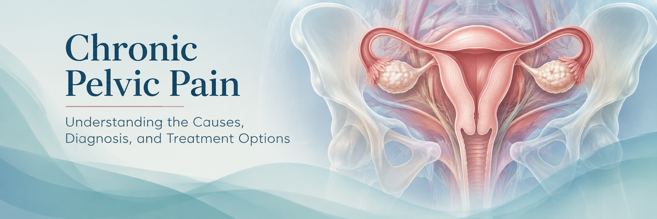
Chronic pelvic pain is pain in the lower abdomen or pelvis that persists or recurs over time, often for six months or longer. It may be constant or intermittent, related to menstruation or intercourse, or associated with urination or bowel movements. For some women, it interferes with work, sleep, relationships, and everyday activities.
Chronic pelvic pain is real, even when examination findings and imaging studies are normal. It is often difficult to diagnose because there may be more than one contributing cause, and some sources of pain cannot be seen on an ultrasound or identified during surgery. Our goal is to listen carefully, look for treatable conditions, and work toward meaningful improvement without unnecessary tests or procedures.
Why Is Chronic Pelvic Pain Difficult to Diagnose?
Several organs, muscles, and nerves share the same region of the pelvis. Pain arising from the uterus, bladder, bowel, pelvic floor muscles, abdominal wall, or nerves may feel very similar. More than one condition can be present at the same time.
In addition, persistent pain can change how the nervous system processes pain signals. This is sometimes called central sensitization. The nervous system becomes more responsive to painful signals, and discomfort may continue even when no ongoing tissue injury is found. This does not mean that the pain is imaginary or psychological.
Possible Causes
Gynecologic conditions
- Endometriosis: Tissue similar to the uterine lining grows outside the uterus and may cause painful periods, intercourse-related pain, or chronic pelvic discomfort. It may be present despite a normal ultrasound.
- Adenomyosis: Endometrial-type tissue within the uterine muscle can contribute to painful, heavy periods and pelvic tenderness.
- Ovarian cysts: Some cysts cause episodic pain, although many small, uncomplicated cysts are incidental findings and do not explain longstanding pain.
- Pelvic inflammatory disease: Infection may cause pelvic pain, and some women experience persistent pain following an infection.
Other common contributors
- Pelvic floor muscle dysfunction: Tight, tender, or poorly coordinated muscles can cause pelvic pressure, pain with intercourse, and pain during examination.
- Bladder pain syndrome: Pain or pressure related to bladder filling, often with urinary frequency or urgency, in the absence of another clear explanation.
- Gastrointestinal conditions: Irritable bowel syndrome, constipation, and other bowel disorders can produce pelvic discomfort.
- Musculoskeletal or nerve-related pain: Abdominal wall trigger points, back or hip disorders, and irritated pelvic nerves can refer pain into the pelvis.
When Should You Seek Urgent Care?
Seek prompt medical attention for sudden or severe pelvic pain, especially with fainting, fever, vomiting, heavy bleeding, or a possible pregnancy. These symptoms may indicate a problem requiring immediate evaluation rather than a routine chronic pain appointment.
How Is Chronic Pelvic Pain Evaluated?
The most useful starting point is often a detailed history. We may ask when the pain began, where it occurs, whether it changes with menstrual cycles, and whether it is associated with intercourse, urination, bowel movements, movement, or previous surgery.
A focused examination may include the abdomen, pelvic organs, and pelvic floor muscles, depending on symptoms and comfort. Tests are selected according to the findings rather than ordered routinely for every patient.
- Pelvic ultrasound: Can identify fibroids, some ovarian cysts, and other structural abnormalities, but does not rule out endometriosis or pelvic floor pain.
- Urine and infection testing: May be appropriate when urinary symptoms or risk factors for infection are present.
- Additional imaging or specialist evaluation: Occasionally needed when bowel, bladder, musculoskeletal, or other causes are suspected.
- Laparoscopy: May be considered in selected cases, particularly when endometriosis is suspected and treatment decisions would be affected. It is not required for every patient and may not reveal the cause of pain.
What Treatments May Help?
There is no single treatment that works for every woman. Treatment depends on the likely causes, examination findings, prior therapies, reproductive plans, and personal goals.
- Hormonal treatment: Continuous hormonal contraception or other menstrual suppression may help pain associated with periods or suspected endometriosis.
- Pelvic floor physical therapy: Can be especially valuable when pelvic muscles are tender, tense, or contributing to pain with intercourse.
- Treatment of bladder or bowel disorders: May substantially improve symptoms when these systems contribute to the pain.
- Pain-management strategies: Selected medications, movement-based rehabilitation, sleep improvement, and behavioral pain therapies can help reduce pain-related disability. These approaches address pain processing and coping, not an assumption that symptoms are imagined.
- Surgery: May be helpful for a clearly identified surgical condition, but is not a reliable treatment for unexplained chronic pelvic pain. Hysterectomy does not guarantee relief, particularly when pain has multiple causes.
What If All the Tests Are Normal?
Normal tests can be reassuring because they make some serious structural conditions less likely, but they do not mean that the pain is unimportant. Conditions involving pelvic floor muscles, nerves, bladder function, or pain processing may not appear on standard imaging. A normal workup should lead to a reassessment of possible contributors and a practical treatment plan—not dismissal of symptoms.
Our Approach
We recognize that chronic pelvic pain can be exhausting and discouraging. We aim to identify treatable causes, explain what testing can and cannot show, and avoid procedures unlikely to help. When appropriate, care may involve pelvic floor physical therapy or collaboration with other specialists. Improvement may be gradual, and success can mean less pain, better daily function, and a better quality of life.
References and Further Reading
- American College of Obstetricians and Gynecologists. Chronic Pelvic Pain. Practice Bulletin No. 218. Obstet Gynecol. 2020;135:e98–e109. doi: 10.1097/AOG.0000000000003716.
- American College of Obstetricians and Gynecologists. Chronic Pelvic Pain. Patient FAQ. ACOG patient information.
- European Association of Urology. EAU Guidelines on Chronic Pelvic Pain. Guideline overview.
- National Institute for Health and Care Excellence. Endometriosis: diagnosis and management (NG73). Clinical guidance.